
How to Choose the Best Fracture Fixation Methods?
Choosing the best fracture fixation methods is a clinical decision, not a simple product comparison. The right method must restore alignment, provide stability, and support safe healing. It should also respect soft-tissue condition, bone quality, fracture location, and the patient’s daily demands.
A displaced wrist fracture may need a plate, while a long-bone fracture may require intramedullary nailing. External fixation can offer valuable temporary stability when swelling or open wounds make internal surgery unsafe. These choices depend on imaging, fracture pattern, circulation, infection risk, and the surgeon’s experience. Patient age, activity level, medical conditions, and willingness to follow rehabilitation plans also matter.
Small details can change the plan. A thin layer of damaged skin may limit incision placement. Poor bone density may reduce screw purchase. A fracture extending into a joint may require especially accurate reduction. No implant is automatically best. Sometimes the safest option is staged treatment, with reassessment after swelling decreases.
Evidence-based guidelines provide a strong foundation, but they cannot replace careful examination. Even experienced teams may revise their strategy during surgery. That is not necessarily failure; it reflects changing information. Still, every decision should have a clear rationale, documented risks, and realistic recovery expectations. This guide examines common fracture fixation methods and the factors that help clinicians choose among them. Final treatment decisions must come from a qualified orthopedic professional after reviewing the patient’s complete clinical picture.
Define Fracture Pattern with AO/OTA Classification and Stability Scores
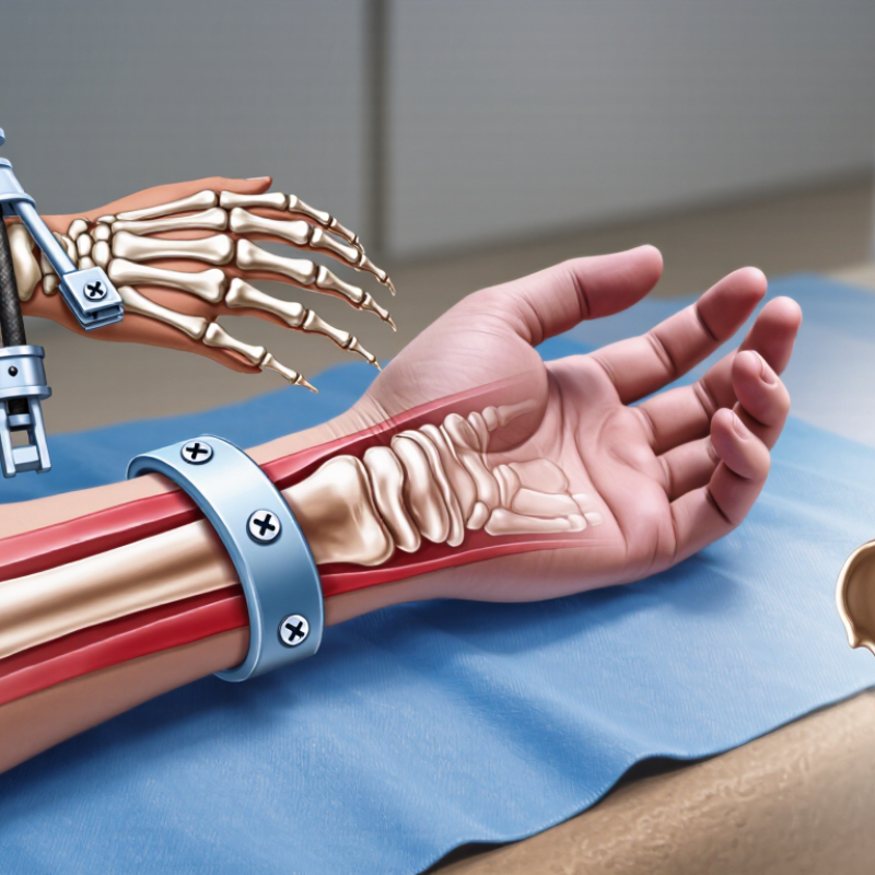
Choosing fracture fixation begins with a disciplined reading of the fracture pattern. The AO/OTA Classification Compendium 2018 separates injuries by bone, segment, morphology, and complexity. This language converts a radiograph into a reproducible description. It also supports clearer communication between surgeons, radiologists, and rehabilitation teams. Yet classification is not perfectly objective. Interobserver agreement can remain moderate, especially with subtle joint involvement or multifragmentary patterns.
Stability scores add clinical meaning beyond the classification code. Assess displacement, comminution, cortical contact, bone quality, soft-tissue damage, and expected loading. A simple fracture with good cortical contact may tolerate controlled motion. A comminuted fracture with poor support may require stronger fixation and slower progression. Do not treat the score as an automatic answer. It is a structured estimate, not a substitute for judgment.
The Global Burden of Disease 2019 study estimated 178 million new fractures worldwide in 2019. That scale makes consistent decision-making increasingly important. In practice, I recheck the score after reviewing computed tomography and soft-tissue findings. I have sometimes underestimated instability when the main fragments looked aligned. Small details matter. A classification can describe the injury, but patient age, activity, healing potential, and surgical risk still influence fixation choice. The framework is useful, although it needs reflection before it becomes a plan.

Screen Patient Risks Using AAOS Data on 300,000 Annual U.S. Hip Fractures
How to Choose the Best Fracture Fixation Methods?
AAOS educational data indicate that about 300,000 older adults in the United States experience hip fractures annually. That number reflects more than surgical demand. It signals a large, medically diverse population requiring careful risk screening. Before selecting fixation, assess fracture location, displacement, bone density, mobility, cognition, and baseline independence. Review anticoagulants, kidney function, heart disease, and infection risks. A stable fracture may suit screws or a sliding device. Unstable patterns may require a stronger intramedullary option. Severe joint damage can change the discussion toward replacement rather than fixation. The fracture pattern decides more than habit.
Tips: Match the method to the patient, not only the X-ray. Confirm imaging in multiple views. Document walking ability before injury. Discuss blood loss, reoperation, weight-bearing limits, and rehabilitation. Ask whether the patient can follow restrictions at home. Small details matter.
AAOS figures help clinicians understand the scale of hip fractures, but population data cannot replace bedside judgment. A 300,000-case estimate does not predict one person’s healing. Frailty, bone quality, and support after discharge can alter outcomes sharply. In practice, the “best” method may be the one that permits safe mobilization with acceptable surgical stress. This is not always obvious. Even experienced teams should reassess the plan when new imaging or medical findings appear. Careful consent should explain uncertainty, expected recovery, and possible complications in plain language.
How to Choose the Best Fracture Fixation Methods?
Screen Patient Risks Using AAOS Data on 300,000 Annual U.S. Hip Fractures
Approximately 300,000 older adults are hospitalized for hip fractures in the United States each year. About 90% are related to falls, roughly 25% of patients die within one year, and approximately 50% do not regain their previous ability to walk independently. These signals support individualized fixation planning based on fall mechanism, frailty, mobility, bone quality, and overall medical risk.
Sources: American Academy of Orthopaedic Surgeons (AAOS), “Hip Fractures”; National Institute on Aging, “Hip Fractures.”
Compare Plates, Nails, and External Fixators by Union and Infection Rates
How to Choose the Best Fracture Fixation Methods?
Union and infection rates help compare plates, intramedullary nails, and external fixators. They should not replace clinical judgment. Cochrane reviews of tibial shaft fractures generally report similar union outcomes for nails and plates, often around 90% to 95%. Intramedullary nails may reduce wound complications, especially when soft tissue damage is significant. However, anterior knee pain remains a practical concern after nailing. Small details matter.
External fixation can protect severely injured skin and muscle without extensive surgical exposure. Its weakness is pin-site infection. Orthopaedic trauma studies commonly report pin-track infection rates between 20% and 40%, although deep infection is less frequent. Union rates often fall near 75% to 90%, depending on fracture type and frame management. Plate fixation may achieve reliable alignment, but infection risks rise when coverage is poor. Reported deep infection rates commonly range from 3% to 10%.
These figures come from systematic reviews, registry analyses, and professional trauma literature, including Cochrane evidence and Orthopaedic Trauma Association publications. The populations differ. That limits direct comparison. A clean, closed fracture in a healthy adult is not equivalent to a contaminated open fracture. Smoking, diabetes, bone loss, injury energy, and surgical timing can change outcomes sharply. I would treat every percentage as a guide, not a promise. Even experienced teams can face delayed union or infection. The best method balances biology, stability, soft-tissue condition, and the patient’s ability to attend follow-up.
Select Open-Fracture Fixation Using BOAST Guidelines and Infection Evidence
Choosing open-fracture fixation requires more than selecting the strongest implant.
BOAST 4 stresses urgent assessment, intravenous antibiotics, vascular review, and coordinated orthoplastic care. Antibiotics should be given ideally within one hour of injury. The wound needs sterile coverage, not repeated emergency inspection.
Fixation should match contamination, bone loss, soft-tissue condition, and patient stability. Temporary external fixation can protect alignment when swelling or contamination remains significant. Definitive internal fixation is appropriate when stable soft-tissue cover can be achieved. BOAST advises that fixation and coverage should occur together whenever possible. A clean-looking wound can still hide devitalised tissue.
Evidence also challenges routine surgical habits.
The FLOW trial included 2,408 patients with open fractures. High-pressure irrigation did not significantly reduce reoperation compared with low-pressure irrigation. Excessive pressure may damage tissue. More treatment is not always better.
Reported infection risk varies sharply by injury severity. A systematic review found infection rates near 1% in lower-grade open fractures, but substantially higher rates in severe grade III injuries. These figures are useful, though they cannot replace individual assessment. Evidence is imperfect. Gustilo classification can vary between observers, and early grading may be wrong.
The fixation plan should remain adjustable. Reassess perfusion, swelling, wound viability, and contamination before committing to definitive hardware. Document antibiotic timing and multidisciplinary decisions clearly. Small delays can matter. A technically successful fixation may still fail when coverage is late or inadequate.
Sources: BOAST 4, Open Fractures, British Orthopaedic Association; FLOW Trial, New England Journal of Medicine, 2015; systematic infection-rate reviews of open fractures.
Verify Healing with Radiographic Union, Reoperation, and Functional Outcomes
Choosing a fracture fixation method should begin with the patient, not the device. Fracture location, bone quality, soft-tissue damage, age, and activity goals influence stability. A technically strong operation can still fail when follow-up is weak. That lesson is easy to underestimate. Clinicians should define healing measures before surgery. Radiographic union is one essential marker. Repeated images can show bridging bone, maintained alignment, and hardware-related changes. Yet an X-ray is not the whole recovery.
At scheduled visits, compare pain, tenderness, walking tolerance, grip strength, or joint motion with baseline findings. Functional outcomes reveal whether the repair works in daily life. A patient may show bridging bone but still struggle with stairs or lifting a cup. Validated questionnaires improve consistency, although answers can vary with mood and expectations. Reoperation rates add another reality check. Record infection treatment, loss of alignment, delayed union, and implant removal separately. Do not label every second operation a failure; some procedures are planned after healing. The context matters.
Reliable assessment needs clear timelines and complete records. Radiographic union should be interpreted with symptoms and examination findings, not alone. Surgeons may review healing at six weeks, three months, and later when needed, adjusting for fracture type. These intervals are not universal. Clinical review often shows that missing one follow-up image creates false confidence. A delayed scan may hide slow progress, while early union may not predict function. Patient-reported limitations should remain visible in the record.
Related Posts
-
10 Trauma Surgery Tips for Global Medical Buyers
-
2026 Top Trends in Orthopedic Implants for Better Patient Outcomes?
-
7 Tips for Choosing Minimally Invasive Spine Surgery
-
China Top Innovations in Bone Regeneration Medicine for Better Health?
-
What Are the Top Stages of the Bone Healing Process?
-
China Best Orthopedic Rehabilitation Programs for Effective Recovery?